Multi-Epitope-Ligand-Cartography (MELC)
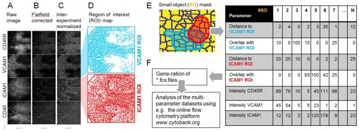
MELC is a massive multiparameter microscopy approach based on fluorescence staining, imaging and bleaching cycles of up to 50 epitopes in the same tissue site. In the course of Project Z01 of the SFB 854, we have established antibody panels to detect all major immune cell subsets in PFA-fixed tissue samples from different organs. For efficient analysis, we developed a workflow, which allows a combined exploration of segmented objects (e.g. cells, but also small random particles) with related fluorescence intensities and spatial distributions using flow cytometry analysis tools. This allows not only the analysis of marker combinations on cells in a specific tissue, but also their spatial relationship, i.e. recruitment to specific tissue zones and interactions between different cell types. Sample preparation and choice of antibody panels for MELC analyses is planned in close collaboration with platform specialists. After the MELC run, the image data is processed and analyzed with the support of the platform specialists.

Figure: Exemplary MELC analysis strategy for leukocyte localization relative to ICAM1- and VCAM1-positive regions of interest (ROI) in the spleen. (A) A series of raw fluorescence images by MELC. The corresponding phase contrast images (not shown) were used to align all images of the image series. (B) Illumination faults were corrected using a flat-field image. (C) Fluorescence intensity images after inter-experiment normalization. (D) Regions of interest defined by using an automated threshold based algorithm. (E) A given field of view is broken down into a mask of randomly distributed “small objects” (SO, yellow) 10 pixels each. For all SO minimal distances to reference regions (e.g. VCAM1-rich in light blue or ICAM1-rich in red) or the pixel overlap of different markers can be determined. (F) Spatial data as well as the fluorescence data for all SO are stored into FCS 3.0 data files and uploaded to the online multi-parameter flow cytometry data analysis platform www.cytobank.org.


